Report
Unknown Substance Found on Spotted Cucumber Beetle Larva (Diabrotica undecimpunctata howardi Barber) [pdf]
Fleming, D. E.
Department of Entomology and Plant Pathology, Mississippi State University, Box 9775, Mississippi State, MS 39762. def18@msstate.edu
Accepted: 27-I-2009
Case Study
An unknown substance (Figure 1) was discovered on the cuticle of a one-day-old spotted cucumber beetle larvae (Diabrotica undecimpunctata howardi) by means of scanning electron microscopy. The larva was from a colony housed in the Insect Rearing Center at Mississippi State University. Larvae were crawling on muslin cloth that serves as a substrate for the eggs until hatch. Samples of larvae were collected and placed in 70% ethanol until they could be prepared for microscopy. Samples were then further dehydrated in an ethanolic series solution to 100% ethanol, critical-point dried in a Polaron E 3000 critical-point drier (Quorum Technologies, Newhaven, UK), mounted on aluminum stubs, and sputter coated with gold-palladium. Digital images of the specimens were acquired by using a JEOL JSM-6500 FE Scanning Electron Microscope (JEOL USA, Peabody, MA).
Suggestions from entomology and plant pathology students and professors, bacteriologists, and virologists have included: wax, fungus, bacterium, protein, and lipid. However, the identity of the substance is still unknown. This substance has an enlarged globular base, and usually has a spiral tail that originates from the center of the base. The substance extensively covered the cuticle (Figure 1). Interestingly, the spirals seem to have a right-hand twist in some cases, but a left-hand twist in others (Figure 2, arrows A and B), indicating this is a non-living substance, as the handedness of living spirals are genetically determined (Peterson 2008). Also, there is an indication that the substance is hollow, due to an area that appears to have collapsed at the end of one of the spiral tails (Figure 2, arrow C). Pores in the insect cuticle can be seen, and in some cases the substance was possibly being exuded from these pores (Figure 2 arrow D). Figure 3 shows a peg possibly “sprouting” on one of the bases, which could be the beginnings of a spiral tail. Figure 4 shows globular bases that are possibly in the process of cleaving or coalescing. Figure 5 shows a peg on the posterior of bases that apparently have been released from the pores. Figure 6 shows the unknown substance on a seta. Other larvae were examined from other batches, and this substance was not found. A search of the literature has revealed very little to lead the author to conclude that the substance is any one of the above suggestions. The author asks that if anyone knows what this substance is to contact him at def18@msstate.edu, or to contact Jack Reed at jreed@entomology.msstate.edu.
Acknowledgements
The author thanks everyone who has given their suggestions and to William Monroe for specimen preparation and taking the digital images.
References
Peterson, C. J. 2008. Personal communication. October 28, 2008. Chris J. Peterson, Research Entomologist USDA Forest Service; Insects, Diseases and Invasive Plants Research Unit, 201 Lincoln Green, Starkville, MS 39759. phone (662) 325-0199; cjpeterson@fs.fed.us.
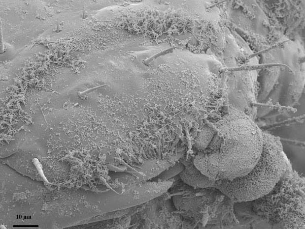
Figure 1. Distribution of the substance on a larval head capsule. Magnification of 950X.
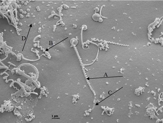
Figure 2. A: a left-hand twist; B: a right-hand twist; C: possible evidence for a hollow tail; and D: cuticular pores and the substance possibly being exuded. Magnification of 6000X.
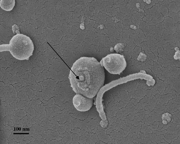
Figure 3. Peg possibly preparing to “sprout” and become a tail. Magnification of 25000X.
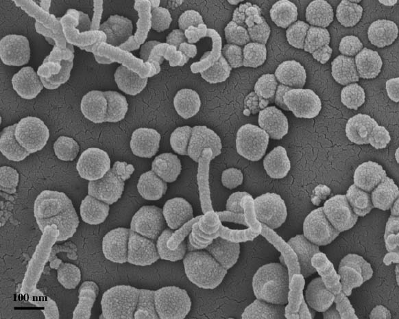
Figure 4. Possible cleaving or coalescing of bases. Magnification of 23000X.
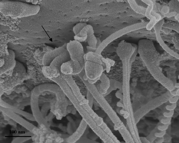
Figure 5. Bases falling from pores, showing peg that might be an attaching mechanism. Magnification of 30000X.
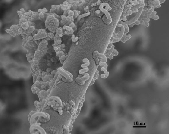
Figure 6. Substance on the surface of seta. Magnification of 23000X.